The Artificial Eye
Reasons for Needing Eye Removal Surgery
There are three main reasons why one’s natural eye needs to be removed.
1. If the natural eye becomes blind and painful. Its removal can give the patient welcome comfort and relief. This is usually only done when most other treatments have failed
2. If the natural eye becomes blind and looks cosmetically poor. For example, the natural eye may look very scarred, start to shrink in size and the cornea (clear window at the front of the eye) may turn white.
3. If the natural eye becomes dangerous e.g. for cancer of the eye
-
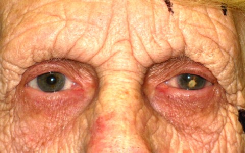
This patient had a blind unsightly left eye which was painful and shrunken
-
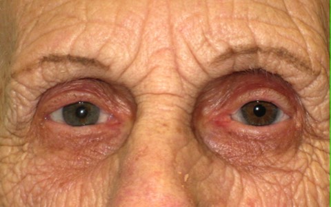
3 months after removal of the left eye and wearing a new artificial eye, the patient is very happy with appearance and comfort
Aims of treatment
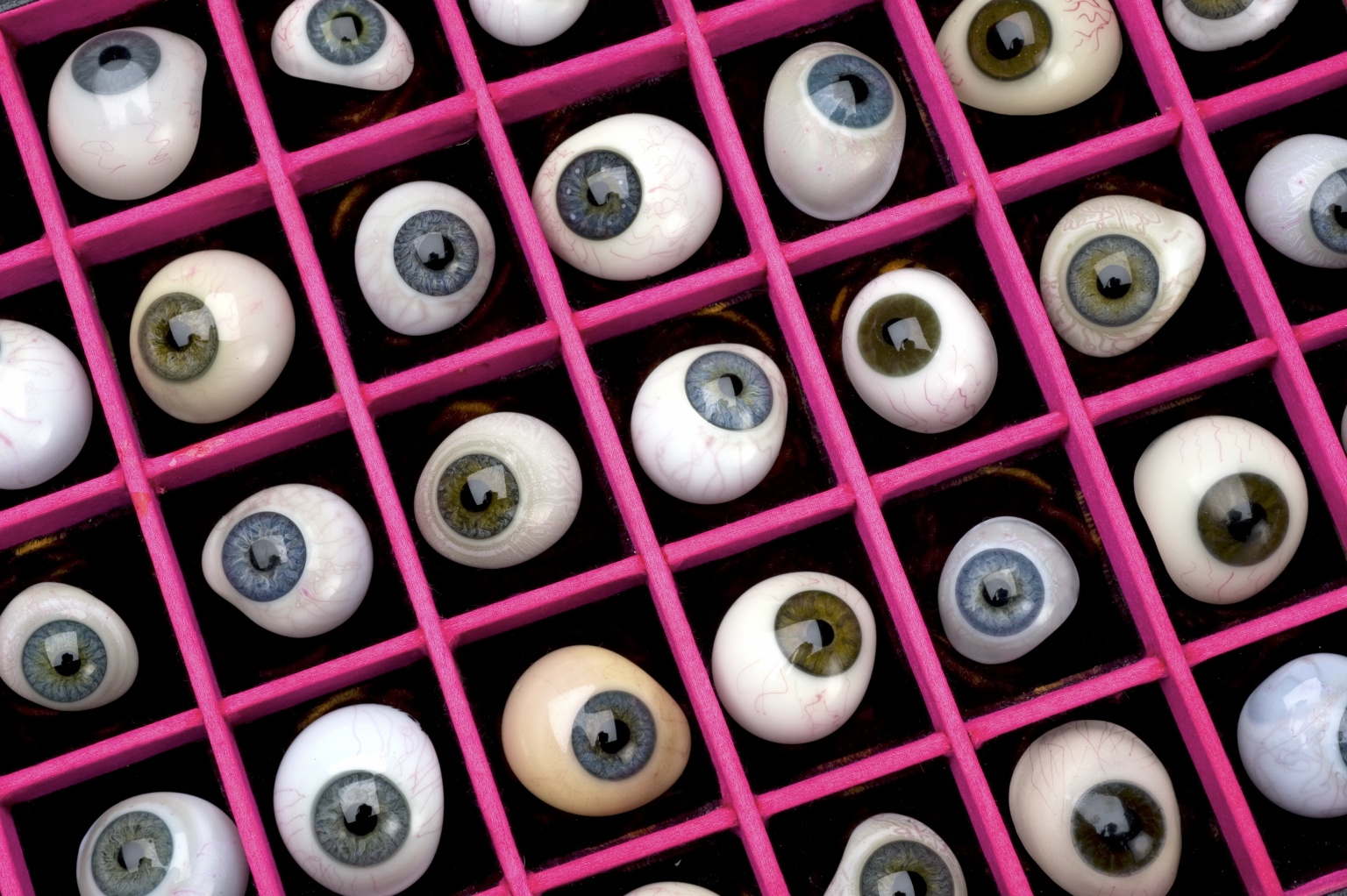
Artificial eyes are custom made in painstaking detail to give an optimum fit and appearance bespoke for that patient
The aims with all artificial eye treatment is to help the patient through the operation of eye removal and to ensure the patient is happy with its appearance, its comfort and the psychology of having an eye socket without a natural eye.
The perfect artificial eye is one which:
• Is comfortable
• Shows good movement
• Is indistinguishable to the observer
• Is easy to look after
The Operation
There are essentially two types of operation for removal of the eye: Evisceration and Enucleation.
Evisceration- involves removal of the clear window at the front of the eye (cornea) & the core of the eye, leaving the outer casing of the eyeball (scleral sac).
Enucleation- involves removal of the entire eyeball.
In both types of operation, an orbital implant which is usually shaped like a ball is inserted deep into the eye socket. The aim of the orbital implant is to fill out the socket, thus replacing some of the lost volume from removed eyeball. Thus the artificial eye (prosthesis), which is later worn by the patient , can be made smaller. The outer coverings of the eyeball (conjunctiva) are then sutured together so that the cavity of the socket is shallower and lined with the pink membrane (conjunctiva). Most patients nowadays undergo an evisceration since it is quicker, thought to offer better motility and has a lower rate of long term problems. However, enucleations are still performed in certain cases such as certain types of eye cancer.
Although both operations are usually performed under general anaesthesia (patient asleep and ventilated), if the anaesthetist considers that general anaesthesia is too dangerous e.g. if the patient has severe lung or heart disease, surgery can be performed quite comfortably under local anaesthesia, often with a mild sedative to relax the patient.
After Surgery
Early period after surgery
A pressure dressing is applied to the socket for the 2-3 days to try to reduce the postoperative swelling. Contrary to belief, usually the patient is very comfortable immediately after surgery. A course of antibiotic tablets is usually prescribed with anti-inflammatory medication (ibruprofen or diclofenac). The patent is then reviewed usually one week after surgery and the dressing is removed. A clear plastic disc called a conformer is often inserted into the cavity of the socket at the time of surgery to help the socket attain the correct shape.
At 6 weeks to 8 weeks, the patient is then usually seen by an ocular prosthetist (sometimes called an ocularist). This is someone who is trained in making artificial eyes. A mould of the cavity of the socket is made so that a new custom made artificial eye to match the patient’s other eye can be manufactured. The manufacture of a new cosmetic shell can take a couple of months and a few visits to the ocular prosthetist since it is a highly skilled and laborious task. Each artificial eye is customised in colour, appearance, shape, thickness for each individual patient. For this reason, the final artificial eye can take up to 3 months to make. However, the patient is often given a temporary artificial from the ocular prosthetist’s supply bank until the final one is ready.
After 3 months
The patient is advised on how to look after the artificial eye and socket by the ocular prosthetist. It is usually advisable to remove the artificial eye from time to time e.g. at night or weekly and to clean it. Your ocular prosthetist will be able to advise you about this. Some patients who are uncomfortable with handling the cosmetic shell, may leave the shell in all the time.
Surgical Risks
The risk of things going wrong with artificial eye surgery is extremely low but as with any type of surgery complications and problems can occur.
Infection
With artificial eye surgery, the most serious complication which can happen early on following surgery is infection. This usually responds very well to antibiotics.
Implant extrusion/exposure
As stated above an orbital implant is usually implanted deep into the socket at the time of removing the eye. The main role of the orbital implant is compensate for the loss in volume from removing the natural eye therefore filling out the socket so that the cavity of the socket is shallower. This means the artificial eye can be thinner and lighter, thus allowing better movement and better comfort for the patient. Very, very rarely, the orbital implant may start become exposed and may even work its way out. This may require further surgery to rectify.
Imperfect appearance/comfort
The vast majority of patients are very happy with the appearance, movement & comfort of their artificial eye. However, a tiny proportion of patients request improved motility, a better appearance or greater comfort. This can often be easily achieved but may require additional surgery.
Delayed Problems of Artificial Eyes
After many, many years of wearing an artificial eye, the patient’s socket may start to alter slightly sometimes resulting in an imperfect appearance, comfort or movement of the artificial eye. For example, the artificial eye may start to look sunken, the upper lid may start to droop (ptosis) or the lower lid may start to sag . If the patient wishes, further procedures may be performed to improve matters.
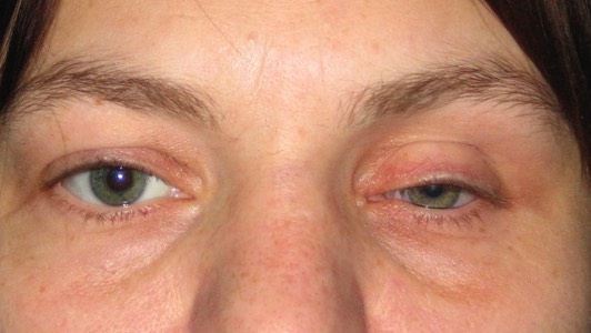
Post enucleation socket syndrome gallery: After many years following eye removal surgery, the artificial eye may sink backwards due to progressive loss of fat from the socket giving the impression of hollowness. Nowadays mild forms of this may be easily treated with hyalauronic acid fillers injected into the eye socket
-
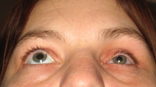
Post Enucleation Socket Syndrome: Before treatment with filler injections. Note how sunken the left (her left) artificial eye has become through many years of wearing an artificial eye.
-
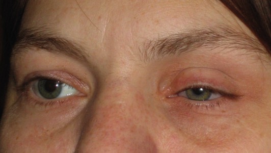
Post Enucleation Socket Syndrome: Before treatment with filler injections. Note how sunken the left (her left) artificial eye has become through many years of wearing an artificial eye.
-
Post Enucleation Socket Syndrome: Before treatment with filler injections. Note how sunken the left (her left) artificial eye has become through many years of wearing an artificial eye.
-
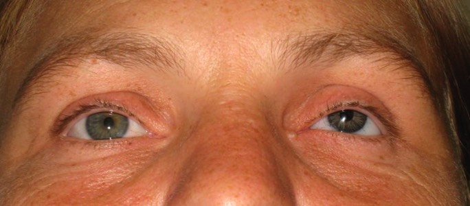
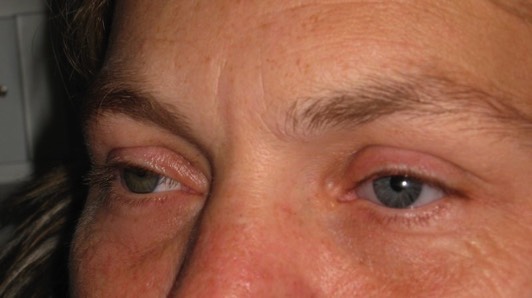
Post Enucleation Socket Syndrome: After treatment with filler injections. Note how this lady's left artificial eye now looks more symmetrical with her right natural eye
-
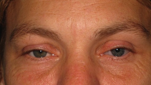
Post Enucleation Socket Syndrome: After treatment with filler injections. Note how this lady's left artificial eye now looks more symmetrical with her right natural eye
-
Post Enucleation Socket Syndrome: After treatment with filler injections. Note how this lady's left artificial eye now looks more symmetrical with her right natural eye