Lump Inside Eyelid I Bumps, Cysts, Styes and Cancers
Introduction
By far the most common lesion of the eyelid is a chalazion, a benign lesion which often self resolves. Despite this, it is important that a qualified doctor with the correct equipment examines the lump to make a proper diagnosis because:
- Some lumps may be cancerous and require early surgery, e.g. Basal Cell Carcinoma. If this is the case, the earlier surgery is performed, the less disfiguring it is likely to be.
- Some lumps may be sight or even life threatening e.g. sebaceous gland carcinoma or squamous cell carcinoma. Prompt treatment may have an effect on improving the patient’s life expectancy.
- Some lumps may indicate a generalised illness. For example Xanthelasma may indicate dangerously high cholesterol levels.
- Some lumps may impair vision. Impairment of vision in young children may lead to poor development of vision and long term vision problems.
Chalazion (Plural: Chalazia) - lump inside eyelid
-
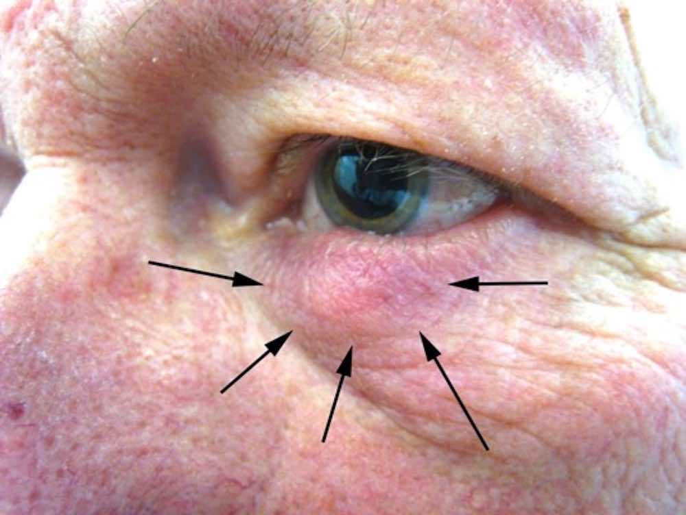
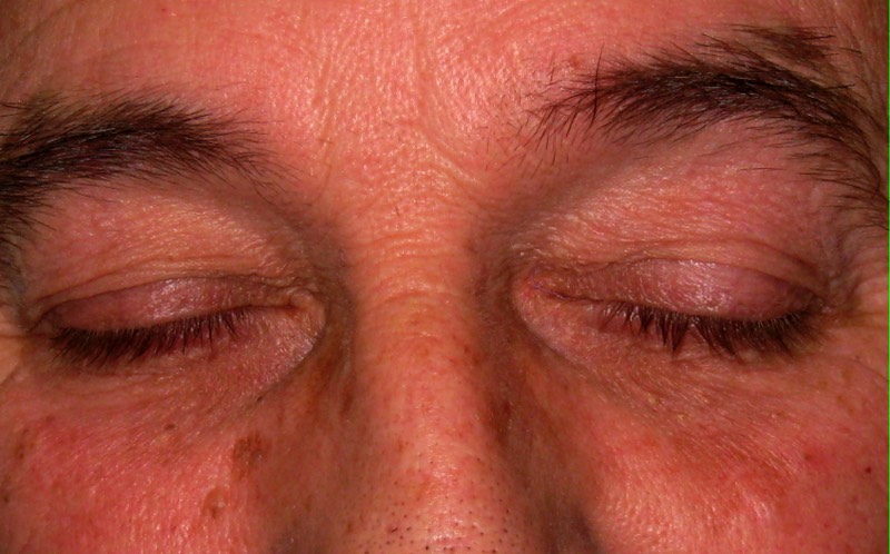
Chalazion: Note the firm swelling of the eyelid. This is occasionally associated with redness and discomfort
-
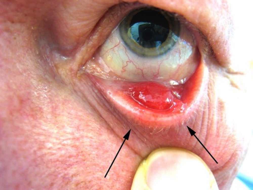
Chalazion: When the eyelid is everted one can see the inflammatory swelling arising from the cartilage like structure on the back of the eyelid
-
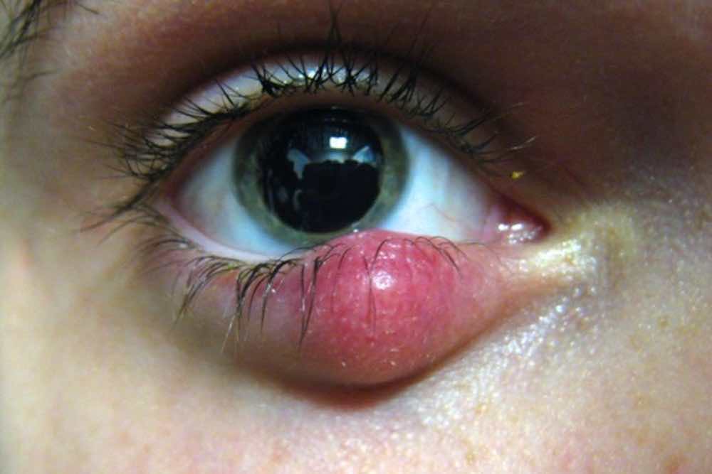
Typical acutely inflamed chalazion. Most chalazia resolve spontaneously although some enlarge so much as to burst either through the front or back of the eyelid
Within the tough cartilage like structure of the back half of the eyelid (known as the tarsal plate) are numerous oil producing glands (meibomian glands) whose job it is to secrete a fine oily secretion on to the surface of the eye to help lubricate it.
Sometimes the duct of a gland can become blocked leading to retention of the oil secretion within the gland. The gland enlarges, as the oil starts to collect and becomes tender as it becomes inflamed.
The patient complains of a red, tender lump of the eyelid. A chalazion, (pronounced KA-LAY-ZEE-ON) also known as meibomian cyst, internal hordeolum represents an inflammatory reaction against the retained oil secretion, (mebum) of the meibomian glands. Eventually the inflammation settles, often resulting in a painless hard lump.
Some patients have an underlying predisposition to chalazion formation due to them also having meibomian gland dysfunction and or blepharitis. The ongoing treatment of MGD is important in reducing the chances of further recurrence of chalazia. (Plural of Chalazion. Pronounced KA-LAY-ZEE-AH)
The patient may notice blurring of vision if the lump is particularly large causing distortion in the normal shape of the cornea.
The vast majority of chalazia will settle down by themselves but hot compresses, using a towel soaked in hot water or proprietary microwaveable beanbags (e.g. MGDRx Bag from the Eyebag Company, Optase bag from Scope Ophthalmics or the Meibopatch Bag from Visufarma) often aids self resolution.
Chalazion & Eyelid Cyst Removal/ Drainage Surgery
Although most chalazia will self resolve in due course, unfortunately a small proportion will remain and can look cosmetically poor. Persistent chalazia can be drained with a simple operation where a small cut is made into the enlarged meibomian gland through the back surface of the eyelid, thus leaving no scar. The procedure is performed under local anaesthetic and all the patient usually feels is the small scratch from the injection. This operation does not unblock the duct orifice of the meibomian gland which caused the chalazion in the first place nor does it cure the patient of any underlying meibomian gland dysfunction; but it does lead to rapid shrinkage of the chalazion.
Unfortunately, rightly or wrongly, funding for the drainage of chalazia has been rationed by the NHS. Mr Cheung's personal opinion is that, although it is understandable that various NHS authorities have rationed the treatment of these lesions, it is important all eyelid lumps are seen by an ophthalmologist and/ or oculoplastic surgeon at least to clarify the diagnosis as every year a small proportion of dangerous eyelid lesions such as BCCs, sebaceous gland carcinomas, are misdiagnosed by the GPs and only referred to ophthalmology services late thus delaying correct treatment.
Stye
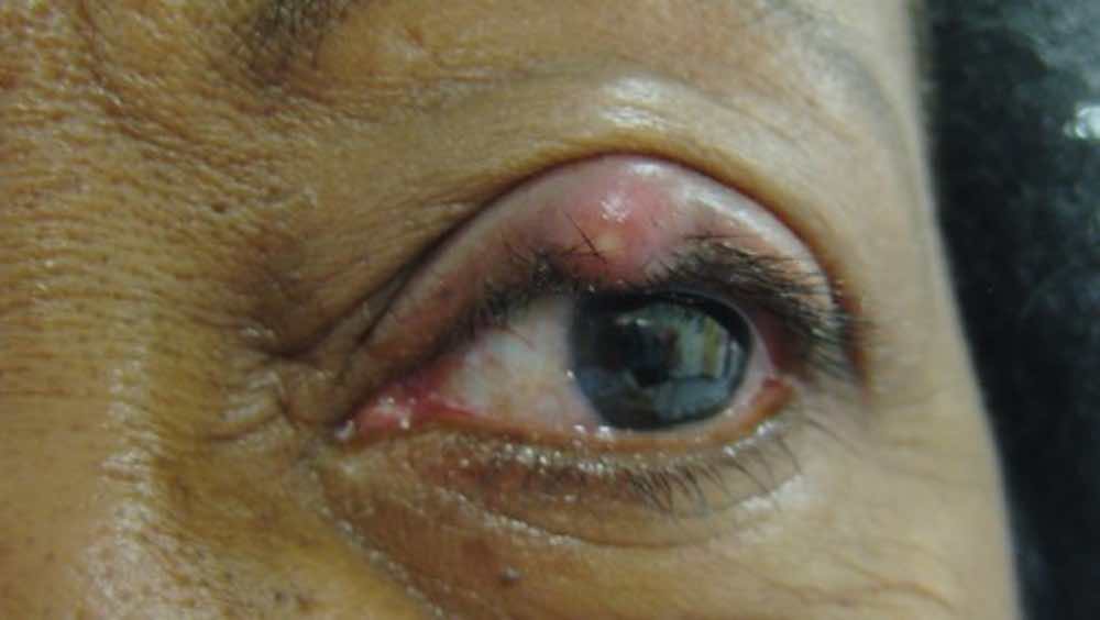
Stye of the upper eyelid. This is due to an acute infection affecting the roots of the eyelashes.
The patient often complains of red tender swelling of the margin of the eyelid. More commonly, styes have a small yellow head of pus.
Like chalazia, hot compresses can help their resolution.
Uncommonly, persistent styes may require surgery which consists of a simple incision into the stye itself under local anaesthesia.
Other Types of Eyelid Lumps, Skin Tags, Milia, Cysts
The skin of the eyelid contains numerous sweat secreting glands and oil secreting glands. Blockage of the openings of these glands leads to the retention of their secretion and the formation of cysts.
- Blocked sweat glands lead to clear fluid containing cysts e.g. Cyst of Moll, Hydrocystoma
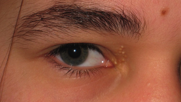
- Blocked oil producing glands lead to white cysts. e.g. cysts of Zeiss
It is however recommended that all patients with any lesion of their eyelid seek a consultation with an ophthalmologist and/ or oculoplastic surgeon as some eyelid cancers may present in a similar fashion.
-
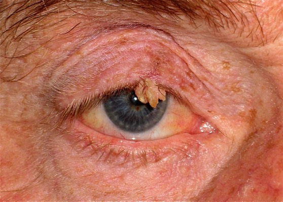
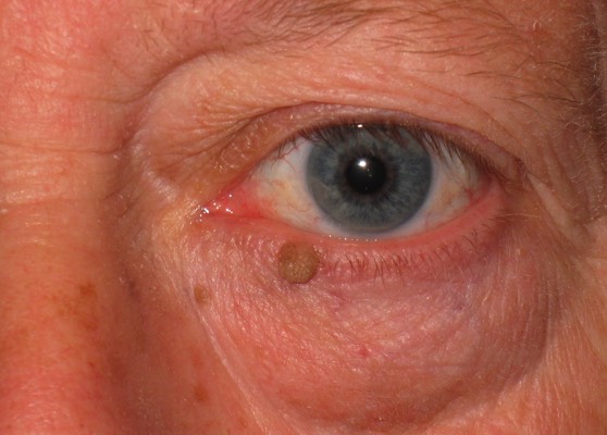
Skin Tag/ Viral Wart: These benign lesions require removal when they obstruct vision.
-
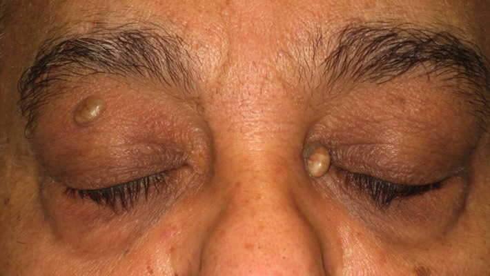
Sebaceous cysts: These white oil filled glands can enlarge to become a cosmetic nuisance
-
Surgery for their removal is quick and often done in the outpatient clinic
-
Some sebaceous cysts can grow quite large. Careful removal is necessary to avoid scarring and collateral damage to local muscles and nerves
-
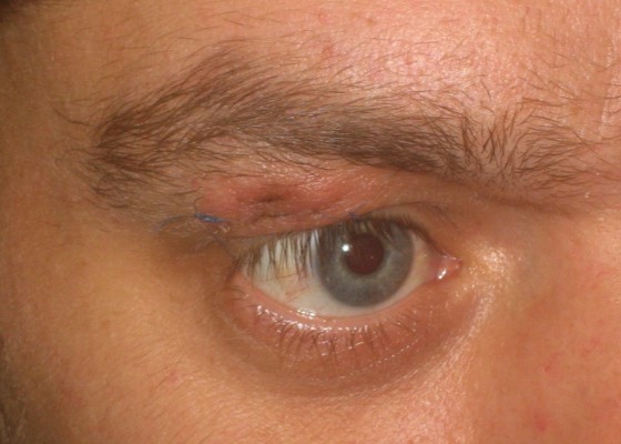
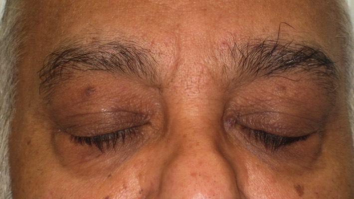
1 week post surgery: The incisions blend away to become invisible.
-
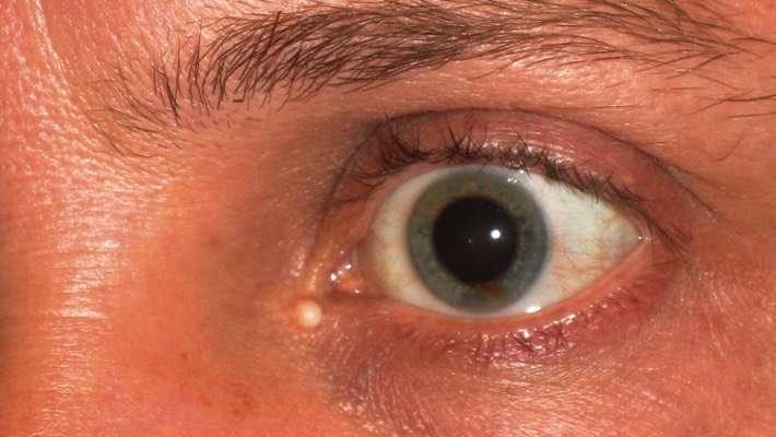
Cyst of Zeiss: Oil filled cyst of the thin skin of the eyelid. Surgery is simple and can be done in an outpatient clinic
-
Skin Tag/ Viral Wart: These lesions are often removed as they represent a cosmetic nuisance.
-
Sebaceous of the eyelids: These are sometimes confused for xanthelasma/ cholesterol patches
-
2 weeks following surgery: Unlike xanthelasma, sebaceous cyst surgery is less complex and less risky.
-
Eyelid Milia: These represent small blockages of the oil producing glands. Surgery often involves simple needle puncture and drainage.
-
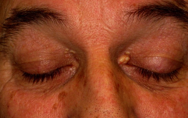
Cyst of Zeiss of the upper eyelid: Oil filled cyst of the thin skin of the eyelid. Surgery is simple and can be done in an outpatient clinic.
-
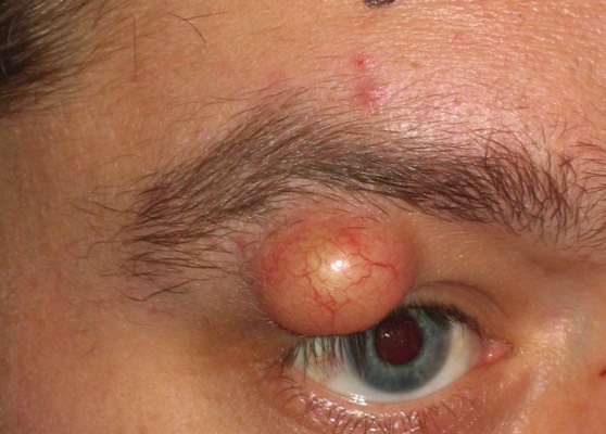
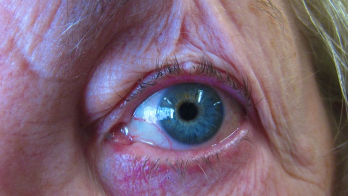
Large cyst of Moll: These represent blockage of the sweat glands. These can enlarge to interfere with function of the eyelid and become quite unsightly
-
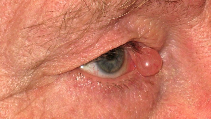
Large cyst of Moll: Surgery for their removal is effective and often done under local anaesthetic. It is important that they be seen by an oculoplastic specialist as some cystic skin cancers can present similarly. Removal for histological analysis is recommended if a benign diagnosis is in doubt.
-
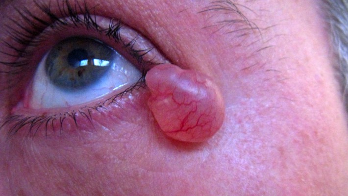
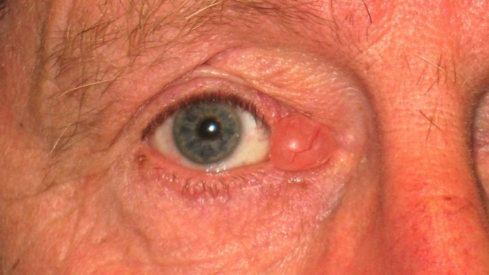
Some benign cysts enlarge to become a functional and cosmetic problem
-
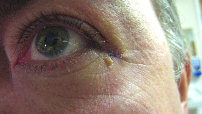
1 week following surgery. The small skin fold smoothes out leaving no trace of surgery nor the original lump.